


Diagnostic Imaging
Nuclear Scintigraphy:
 Nuclear Scintigraphy or “Bone Scanning” is the most commonly performed equine nuclear medicine procedure because it offers the highest sensitivity for early detection of bone disease. In addition, it allows one to evaluate the entire equine skeleton (or a specific region) making it an ideal tool for diagnosing difficult or multiple limb lameness. Nuclear Scintigraphy is an imaging modality that looks at the physiologic process of bone metabolism (how the bone is working). It is especially useful in detecting early bone inflammation, when a lameness is difficult to diagnose or when the horse has poor performance issues that are unexplained. For more information about Nuclear Scintigraphy at Chicago Equine, please click here.
Digital Radiography
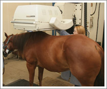
At Chicago Equine we offer digital radiography (DR) both in the field and in hospital. Just as digital cameras have revolutionized photography, DR has changed the face of x-ray technology. We use Eklin DR machines in the field and the new DR system from VetRocket in the hospital. We also have Fuji CR system in the hospital. In less than four seconds, we can take a digital x-ray of the patient’s injury or area of concern without having to waste precious minutes to develop film. The veterinarian can examine, manipulate, and optimize the image, allowing preliminary diagnoses to be made quickly.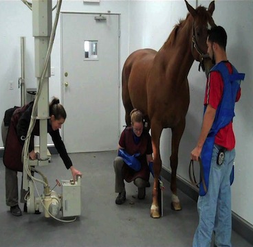
We also use VetRocket Pac system which allows us to integrate any other digital images, such as endoscopy or ultrasound, with the DR images. This helps us evaluate the area of concern more closely and bring additional technologies into our diagnostic process.
Our stationary machine can handle deep tissue penetration and is used primarily for imaging the chest, abdomen, skull, and upper extremities (shoulder, stifle, neck, back, hip, and pelvis).
All of these capabilities help us make a more rapid and confident diagnosis, which means we can start treating the horse sooner. Quicker treatment generally results in a faster and more positive outcome.
Ultrasound
Advances in ultrasonography have changed considerably in the last few years. We can visualize soft tissue structures like never before. The advanced ultrasound machines in use at Chicago Equine provide the highest quality images available. These machines have a Musculoskeletal mode (for tendons, ligaments, muscles, bones, cartilage, etc.) a Nerve mode for visualization of nerves for complicated nerve blocks or nerve root injections, a Cardiac mode, and a General mode for better visualization of the internal organs. We have 6 different probes, which allows us to ultrasound every part of the horse.
Ultrasound is a valuable imaging tool for evaluating and diagnosing many different conditions and monitoring healing progress. Ultrasound uses acoustic waves to produce a real time image of a wide variety of anatomic structures. This modality allows for more accurate detection of minute soft tissue changes and it is more sensitive to changes in bone than radiographs. Ultrasound exams can be routinely performed on the equine athlete to monitor the extent of an injury or to make sure the horse is not going to have a catastrophic injury. Ultrasound exams of the chest and abdomen are performed to diagnose and monitor treatment of diseases including pleuritis, pneumonia, colic, and chronic weight loss. Ultrasound can be used for many procedures including guided biopsies of the liver, kidneys, spleen, lung and other tissues. Ultrasound exam can be used to view the ligaments, bone and articular cartilage of a joint. The most common ultrasound exam is of tendon and ligament injuries in the Sport Horse. An ultrasound exam of the heart is called echocardiography which is used to evaluate the size of the heart, significance of heart murmurs, exercise intolerance and arrhythmias. Ultrasound exams of the spine and pelvis can be performed to diagnose stress fractures, degenerative joint disease as well as guided injections of the dorsal facet joints, sacroiliac joints, and nerve root injections. Ultrasound exams of a brood mare’s reproduction tract can be valuable tool to examine mares to be bred as well as to determine pregnancy. Both hospital and ambulatory veterinarians are equipped with digitally enhanced ultrasound machines.
